Atrophic Pattern On Pap Smear
Atrophic Pattern On Pap Smear - Vaginal atrophy occurs most often after menopause. The test itself involves collection of a sample of cells from a woman's cervix (the end of the uterus that extends into the vagina) during a routine pelvic exam. Ascus may be caused by a vaginal. Human papillomavirus (hpv) according to the centers for disease control and prevention (cdc), the human papillomavirus is a common virus that is transmitted through sexual activity. Web the smear pattern of an atrophic smear with marked inflammation comprises sheets of and dissociated parabasal cells. Web one of the most common abnormal findings on a pap smear —a routine screening test for cervical cancer and any abnormal cell changes on the cervix that might lead to cervical cancer—is known as ascus. It can be done along with a hpv test during a pelvic exam as part of cervical cancer screening for people with no symptoms. In women older than age 30, the pap test may be combined with a test for human papillomavirus (hpv) — a common sexually transmitted infection that can cause cervical cancer. Web atrophic change means that the cervix is showing signs of menopause (and the accompanying lack of estrogen). We are, therefore, primarily interested in detecting any atypical cells. My gyn called & said my pap smear results were atrophic. The virus can cause cell changes that lead to cervical cancer. In liquid based cytology, background of atrophic smear is cleaner. A shortened or narrowed vagina. The hpv test looks for human papillomavirus (hpv). What does atrophic smear mean in pap result. Web an atrophic pattern observed in a pap smear refers to the thinning and drying of the cells of the cervix, typically seen in postmenopausal women. Human papillomavirus (hpv) according to the centers for disease control and prevention (cdc), the human papillomavirus is a common virus that is transmitted through sexual activity. Usual pattern of atrophic smears taking pap smear in postmenopausal women occasionally shows a hypocellular background and may display the absence of endocervical or transformation zone components (6,7) (figure 1a). We are, therefore, primarily interested in detecting any atypical cells. This condition can be caused by hormonal changes during menopause, decreased. Of 67, only 36 cases were positive for hgd with the ppv of only 54% (36/67). Human papillomavirus (hpv) according to the centers for disease control and prevention (cdc), the human papillomavirus is a common virus that is transmitted through sexual activity. What does atrophic smear mean in pap. A doctor has provided 1 answer. For many women, vaginal atrophy not only makes intercourse painful but also leads to distressing urinary symptoms. She stated that this meant that she didn't collect enough cells & i have to have another test? In liquid based cytology, background of atrophic smear is cleaner. Web the smear pattern of an atrophic smear with. You may have to apply the moisturizer every few days. Web the pap test checks for cell changes on a woman’s cervix that could turn into cancer if they are not treated. Web so basically, most women will get two pieces of information: Classic signs of atrophy during a pelvic exam include: Web the interpretation of atrophic vaginitis from papanicolaou. Web vaginal atrophy (atrophic vaginitis) is thinning, drying and inflammation of the vaginal walls that may occur when your body has less estrogen. In women older than age 30, the pap test may be combined with a test for human papillomavirus (hpv) — a common sexually transmitted infection that can cause cervical cancer. We are, therefore, primarily interested in detecting. Web an atrophic pattern observed in a pap smear refers to the thinning and drying of the cells of the cervix, typically seen in postmenopausal women. It can be done along with a hpv test during a pelvic exam as part of cervical cancer screening for people with no symptoms. Human papillomavirus (hpv) according to the centers for disease control. The squamous cells of your cervix were slightly abnormal on your pap smear. Microscopic examination of atrophic smears which is typically seen in postmenopausal women usually shows. Web laboratory diagnostic testing, including serum hormone levels and papanicolaou smear, can confirm the presence of urogenital atrophy (figures 2 and 3; The test itself involves collection of a sample of cells from. Web a healthcare provider can diagnose vaginal atrophy based on your symptoms and a pelvic exam to look at your vagina and cervix. The hpv testing makes the recommendation we give you about how to follow up on your pap result more accurate. My gyn called & said my pap smear results were atrophic. Web so basically, most women will. It can be done along with a hpv test during a pelvic exam as part of cervical cancer screening for people with no symptoms. The test itself involves collection of a sample of cells from a woman's cervix (the end of the uterus that extends into the vagina) during a routine pelvic exam. May resemble urothelial metaplasia, but cells have. Also known as the pap test) is a screening test for cervical cancer. Usual pattern of atrophic smears taking pap smear in postmenopausal women occasionally shows a hypocellular background and may display the absence of endocervical or transformation zone components (6,7) (figure 1a). A shortened or narrowed vagina. The pap smear is usually done in conjunction with a pelvic exam.. Web so basically, most women will get two pieces of information: The hpv test looks for human papillomavirus (hpv). Microscopic examination of atrophic smears which is typically seen in postmenopausal women usually shows. The virus can cause cell changes that lead to cervical cancer. Classic signs of atrophy during a pelvic exam include: Web severe atrophy can show dirty background with inflammation, debris, old blood, blue blobs and giant cells. The pap smear is usually done in conjunction with a pelvic exam. Microscopic examination of atrophic smears which is typically seen in postmenopausal women usually shows. She stated that this meant that she didn't collect enough cells & i have to have another test? 3, 13 cytologic examination of. For many women, vaginal atrophy not only makes intercourse painful but also leads to distressing urinary symptoms. Web the interpretation of atrophic vaginitis from papanicolaou (pap) tests is seemingly subjective, despite descriptive terminology criteria published by the bethesda system for reporting cervical cytology, 1 as evidenced by the historically poor performance on these slides in the college of american pathologists (cap). This condition can be caused by hormonal changes during menopause, decreased. In liquid based cytology, background of atrophic smear is cleaner. What does atrophic smear mean in pap result. Web so basically, most women will get two pieces of information: Of 67, only 36 cases were positive for hgd with the ppv of only 54% (36/67). In women older than age 30, the pap test may be combined with a test for human papillomavirus (hpv) — a common sexually transmitted infection that can cause cervical cancer. Web a pap smear (papanicolau smear; Usual pattern of atrophic smears taking pap smear in postmenopausal women occasionally shows a hypocellular background and may display the absence of endocervical or transformation zone components (6,7) (figure 1a). Web laboratory diagnostic testing, including serum hormone levels and papanicolaou smear, can confirm the presence of urogenital atrophy (figures 2 and 3;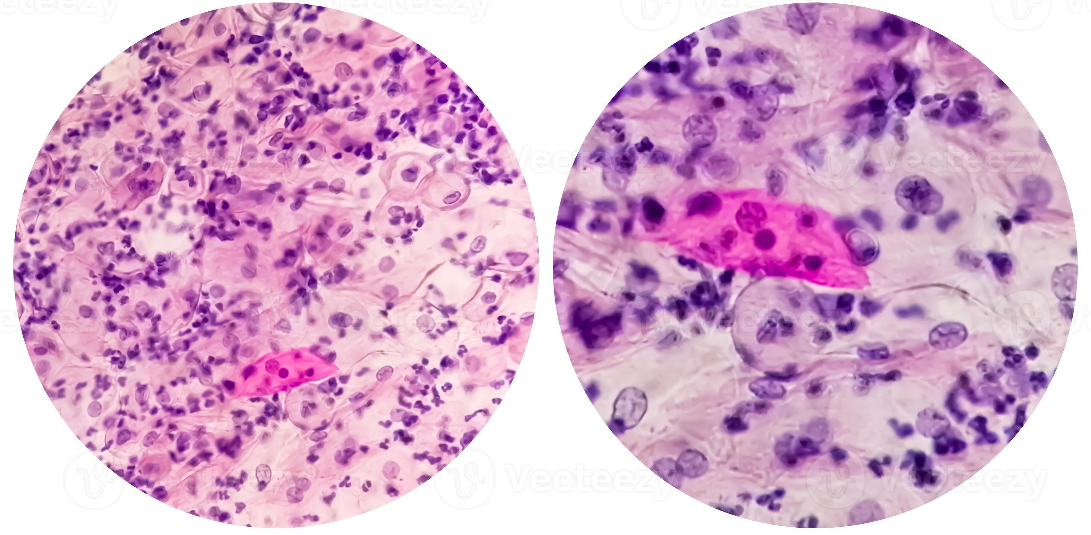
Paps smear. Microscopic examination of pap smear showing inflammatory

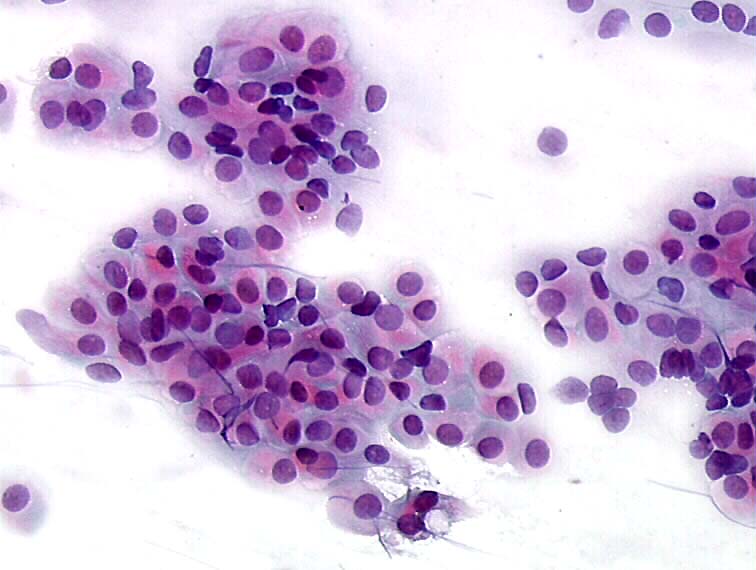
Pap smear cytology showing mostly immature basal cells, typical for
Histopathology and cytopathology of the uterine cervix digital atlas

Paps Smear Microscopic Showing Inflammatory Smear Stock Photo

Conventional smear atrophic smear (Papanicolaou smear, ×200

Premium Photo Paps smear. pap smear showing inflammatory smear with

Understanding The Atrophic Pattern In Pap Smear Results MedShun

Paps smear. Microscopic examination of pap smear showing inflammatory

Paps smear. Microscopic examination of pap smear showing inflammatory

Paps smear. Microscopic examination of pap smear showing inflammatory
May Resemble Urothelial Metaplasia, But Cells Have Prominent Intercellular Bridges.
Loss Of Fragile Cytoplasm Of The Thin Atrophic And Relatively Dry Epithelium Leads To Plenty Bare Nuclei Throughout The Smear.
It Can Be Done Along With A Hpv Test During A Pelvic Exam As Part Of Cervical Cancer Screening For People With No Symptoms.
A Shortened Or Narrowed Vagina.
Related Post: