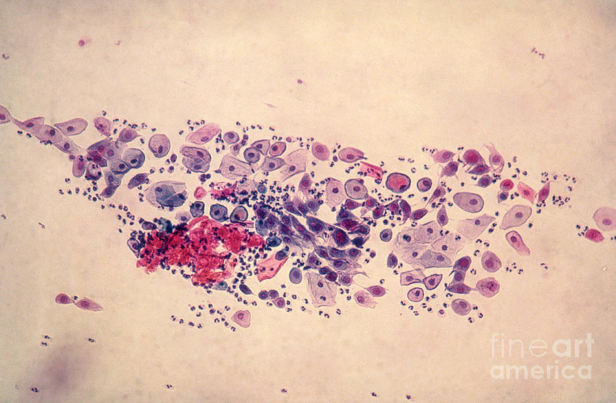
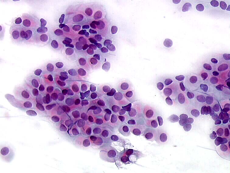
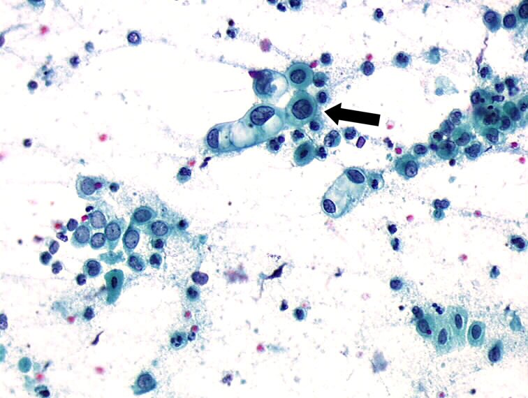
Atrophic Pattern Predominantly Parabasal Cells
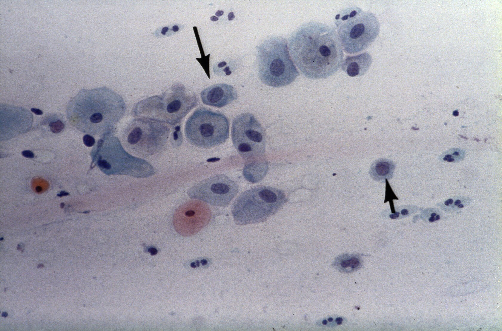
Atrophic Pattern Predominantly Parabasal Cells - An intermediate pattern between the two is also encountered; Web the second is an atrophic pattern that consists predominantly of parabasal cells and is attributed to a lack of estrogenic stimulation (fig. A doctor has provided 1 answer. Web a recognizably atrophic pattern, composed of thick clusters of intermediate and large parabasal cells, was termed “crowded” by koss. Naked nuclei (small cells) may be seen. Web a doctor has provided 1 answer. Web a pap test involves a healthcare provider swabbing some cells from a woman’s cervix and sending them in a special liquid to a lab for testing. The condition also includes urinary tract problems such as urinary tract infections (utis) and urinary incontinence. The hyperchromatic nucleus was relatively round with smooth contours. Web hyperchromatic crowded groups in pap smear with atrophic cellular pattern with occasional atypical degenerated enlarged parabasal nucleus in some of the cells in hyperchromatic crowded groups of parabasal cells. What does this mean ? A doctor has provided 1 answer. Occasionally in our experience, a mixed pattern composed of parabasal, intermediate, and superficial cells can be seen. This is composed predominantly of intermediate cells admixed either with some superficial or parabasal cells. Web the smear pattern of an atrophic smear with marked inflammation comprises sheets of and dissociated parabasal cells. Web vaginal atrophy is a condition where the lining of your vagina gets drier and thinner. Vaginal atrophy occurs most often after menopause. My gyn called & said my pap smear results were atrophic. Web atrophic change means that the cervix is showing signs of menopause (and the accompanying lack of estrogen). Cells have high n/c ratio but uniform chromatin. Web my pap smear (atrophic) shows predominantly parabasal cells with scattered superficial squamous cells. Web what does the result in pap smear c 211 13 and smear 03 mean? My gyn called & said my pap smear results were atrophic. A doctor has provided 1 answer. It only means that the organ less than healthy epithelium, however, if it is. Web vaginal atrophy is a collection of symptoms—including vaginal dryness, dysuria, and vulvovaginal irritation and itching—that are generally associated with declining estrogen levels attributable. We are, therefore, primarily interested in detecting any atypical cells. Web atypical immature metaplasia associated with inflammation and atrophy is a challenge in cervical biopsy interpretation. Naked nuclei (small cells) may be seen. Web the smear. Web your pap test will come back with one of three results: She stated that this meant that she didn't collect enough cells & i have to have another test?: Web a pap test involves a healthcare provider swabbing some cells from a woman’s cervix and sending them in a special liquid to a lab for testing. The hyperchromatic nucleus. Web my pap smear (atrophic) shows predominantly parabasal cells with scattered superficial squamous cells. A doctor has provided 1 answer. Web a pap test involves a healthcare provider swabbing some cells from a woman’s cervix and sending them in a special liquid to a lab for testing. Without the use of estrogen in the vagina or otherwi. However, there are. Naked nuclei (small cells) may be seen. Web vaginal atrophy (atrophic vaginitis) is thinning, drying and inflammation of the vaginal walls that may occur when your body has less estrogen. For many women, vaginal atrophy not only makes intercourse painful but also leads to distressing urinary symptoms. Vaginal atrophy occurs most often after menopause. A doctor has provided 1 answer. This means no cell changes were found. A shift in maturation index in the absence of significant inflammation is more accurately termed atrophic pattern. A doctor has provided 1 answer. She stated that this meant that she didn't collect enough cells & i have to have another test?: Naked nuclei (small cells) may be seen. Vaginal atrophy occurs most often after menopause. This may be accompanied by abundant neutrophils. Web a pap test is a procedure used to collect cells from the cervix (lower part of the uterus) so they can be looked at closely in a lab under a microscope. She stated that this meant that she didn't collect enough cells & i have. However, there are normal to low numbers of neutrophils. Web vaginal atrophy (atrophic vaginitis) is thinning, drying and inflammation of the vaginal walls that may occur when your body has less estrogen. Occasionally in our experience, a mixed pattern composed of parabasal, intermediate, and superficial cells can be seen. A doctor has provided 1 answer. Web vaginal atrophy is a. Web atrophic pattern histologic findings demonstrate decreased superficial squamous cells, increased parabasal cells, decreased lactobacilli. Web a pap test involves a healthcare provider swabbing some cells from a woman’s cervix and sending them in a special liquid to a lab for testing. Vaginal atrophy in menopause shows increased parabasal cells on cytology. A doctor has provided 1 answer. For many. She stated that this meant that she didn't collect enough cells & i have to have another test?: Web the second is an atrophic pattern that consists predominantly of parabasal cells and is attributed to a lack of estrogenic stimulation (fig. My gyn called & said my pap smear results were atrophic. A doctor has provided 1 answer. Occasionally in. Web what does the result in pap smear c 211 13 and smear 03 mean? Vaginal atrophy in menopause shows increased parabasal cells on cytology. For many women, vaginal atrophy not only makes intercourse painful but also leads to distressing urinary symptoms. Vaginal atrophy occurs most often after menopause. Web vaginal atrophy is a condition where the lining of your vagina gets drier and thinner. Naked nuclei (small cells) may be seen. Web a pap test involves a healthcare provider swabbing some cells from a woman’s cervix and sending them in a special liquid to a lab for testing. My gyn called & said my pap smear results were atrophic. Web a recognizably atrophic pattern, composed of thick clusters of intermediate and large parabasal cells, was termed “crowded” by koss. A doctor has provided 1 answer. An intermediate pattern between the two is also encountered; Web a pap test is a procedure used to collect cells from the cervix (lower part of the uterus) so they can be looked at closely in a lab under a microscope. Web atypical squamous cells of undetermined significance (ascus) is the most common abnormal finding from a pap smear. Web my pap smear (atrophic) shows predominantly parabasal cells with scattered superficial squamous cells. A shift in maturation index in the absence of significant inflammation is more accurately termed atrophic pattern. It means that some of the cells from a pap smear did not look entirely normal but did not meet the diagnostic criteria for a lesion (meaning an area of abnormal tissue).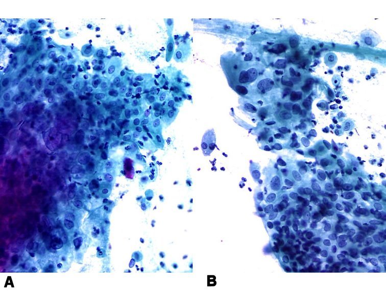
Cytopathology of the uterine cervix digital atlas
Pap Smear, Parabasal Cells Photograph by Science Source

Cytopathology of the uterine cervix digital atlas
Histopathology and cytopathology of the uterine cervix digital atlas

Parabasal cells in pap smear with postpartum Ad , ad, cells

Cell Atrophy

The classi¯cation of cytologic examination. (A) Parabasal cell. (B
Cytopathology of the uterine cervix digital atlas

Cytopathology of the uterine cervix digital atlas
Parabasal cells Collection
Often, An Examination Under The Microscope May Diagnose Inflammations From Several Microorganisms (Bacteria, Fungi, Trichomoniasis, Etc).
Loss Of Fragile Cytoplasm Of The Thin Atrophic And Relatively Dry Epithelium Leads To Plenty Bare Nuclei Throughout The Smear.
This Results In Itching, Burning And Pain During Sex, Among Other Symptoms.
Web A Doctor Has Provided 1 Answer.
Related Post: