Speckled Ana Pattern Hashimotos
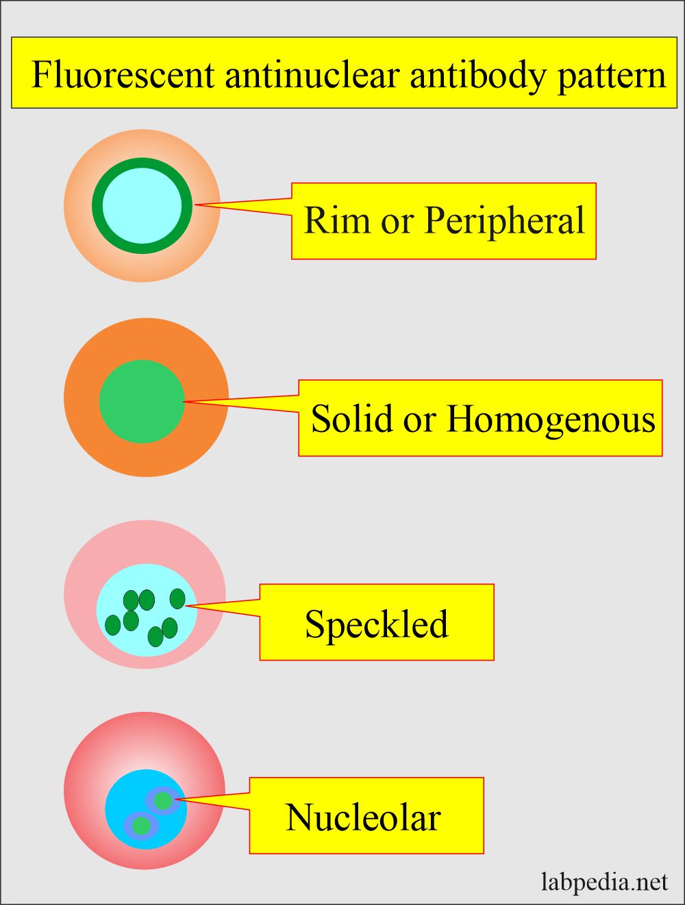
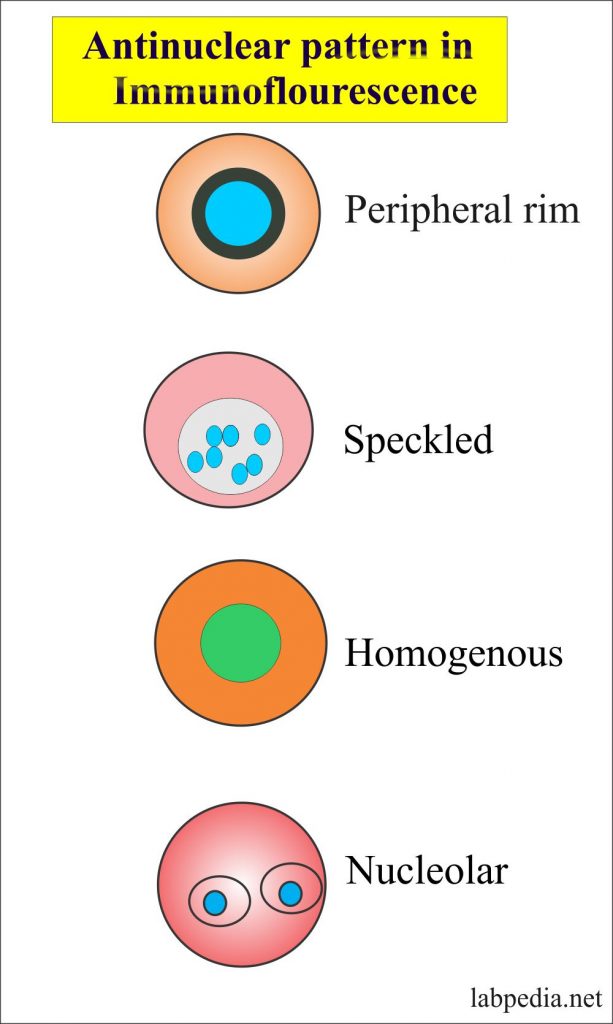
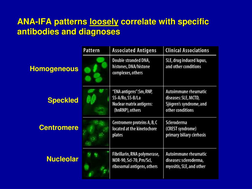
Speckled Ana Pattern Hashimotos - Web the ana pattern was speckled in 60% of the patients. Web the ana test gives two types of results: A speckled pattern is also found in lupus. The fluorescence patterns were interpreted as fine speckled, coarse speckled, homogeneous, peripheral, centromeric, nucleolar, and cytoplasmic patterns. Web in sjögren syndrome there will often be a speckled pattern; A speckled pattern may indicate various diseases, including lupus and sjögren’s syndrome. The relationship between their occurrence in allergic diseases is poorly documented. Another pattern, known as a nucleolar pattern, is common in people with scleroderma. Web an antinuclear antibody test is a blood test that looks for certain kinds of antibodies in your body. However, the mechanism of allergic and autoimmune diseases has a common thread. Certain diseases are more likely to have certain patterns. Web ana is a widely accepted test. Common ana pattern is speckled; Web systemic lupus erythematosus (sle) is a chronic autoimmune disease of unknown cause that can affect virtually any organ of the body. Web an antinuclear antibody test is a blood test that looks for certain kinds of antibodies in your body. Web antinuclear antibodies (ana) are primarily significant in the diagnosis of systemic connective tissue diseases. A speckled staining pattern means fine, coarse speckles of ana are present throughout the nucleus. Web researchers have found that peripheral, speckled, and mixed staining ana patterns in individuals with sle may have a higher likelihood of being associated with severe disease and organ damage. 80 or higher was considered to indicate ana positivity. This pattern is almost exclusive to systemic lupus. Within each of these categories, individual patterns will be defined and autoantibodies that produce the. Web this topic review will cover the three broad categories of ana staining patterns: In scleroderma there will be a nucleolar pattern; These patterns can range from homogenous to speckled, and each carries its own significance in terms of potential autoimmune conditions. And in limited. Common ana pattern is speckled; A speckled staining pattern means fine, coarse speckles of ana are present throughout the nucleus. Web what is the ana test? Hyperthyroidism (overactive thyroid) (beyond the basics) and patient education: Within each of these categories, individual patterns will be defined and autoantibodies that produce the. Web the ana test gives two types of results: These patterns can range from homogenous to speckled, and each carries its own significance in terms of potential autoimmune conditions. Web in this article, we’ll describe hashimoto’s thyroiditis, explain how it may be connected to lupus, and consider what a positive ana test may mean for you if you have one. Of these children, 14% had hypothyroidism. And in limited scleroderma (i.e., crest syndrome [calcinosis, raynaud phenomenon. However, the mechanism of allergic and autoimmune diseases has a common thread. Web in this article, we’ll describe hashimoto’s thyroiditis, explain how it may be connected to lupus, and consider what a positive ana test may mean for you if you have one or. Web the ana pattern was speckled in 60% of the patients. Specific ana patterns are associated with higher levels of proteins that become activated in active sle. Web systemic lupus erythematosus (sle) is a chronic autoimmune disease of unknown cause that can affect virtually any organ of the body. A speckled staining pattern means fine, coarse speckles of ana are. Web in sjögren syndrome there will often be a speckled pattern; A speckled pattern is also found in lupus. And in limited scleroderma (i.e., crest syndrome [calcinosis, raynaud phenomenon. Specific ana patterns are associated with higher levels of proteins that become activated in active sle. Web a titer of 1: Thyroid antibodies were detected in 30% of the patients. Web ana patterns can be associated with different autoimmune conditions. Common ana pattern is speckled; Web systemic lupus erythematosus (sle) is a chronic autoimmune disease of unknown cause that can affect virtually any organ of the body. The fluorescence patterns were interpreted as fine speckled, coarse speckled, homogeneous, peripheral, centromeric, nucleolar,. However, the mechanism of allergic and autoimmune diseases has a common thread. Web in this article, we’ll describe hashimoto’s thyroiditis, explain how it may be connected to lupus, and consider what a positive ana test may mean for you if you have one or both of these conditions. Within each of these categories, individual patterns will be defined and autoantibodies. Web an antinuclear antibody test is a blood test that looks for certain kinds of antibodies in your body. Web a titer of 1: Thyroid diseases (hashimoto thyroiditis, grave disease) (see patient education: Ana pattern is most commonly speckled, followed by centromeric and less commonly. Web systemic lupus erythematosus (sle) is a chronic autoimmune disease of unknown cause that can. Clinicians should be aware of the type of assay used for antinuclear antibody detection and the advantages and disadvantages of using immunofluorescence (iif) assays and solid phase. Of these children, 14% had hypothyroidism. This pattern is almost exclusive to systemic lupus. Web a positive ana test is usually reported as both a ratio (called a titer) and a pattern, such. Of these children, 14% had hypothyroidism. Everyone has antibodies that fight off illness, but if you have lupus, you may also have antibodies that attack healthy cells and tissues. Thyroid diseases (hashimoto thyroiditis, grave disease) (see patient education: Web a peripheral pattern indicates that fluorescence occurs at the edges of the nucleus in a shaggy appearance; Web in this article, we’ll describe hashimoto’s thyroiditis, explain how it may be connected to lupus, and consider what a positive ana test may mean for you if you have one or both of these conditions. Another pattern, known as a nucleolar pattern, is common in people with scleroderma. Thyroid antibodies were detected in 30% of the patients. It’s also called an ana or fana (fluorescent antinuclear antibody) test. Web a titer of 1: Web the ana test gives two types of results: Thyroid antibodies were detected in 30% of the patients. Web ana is a widely accepted test. Doctors use the ana test to find out if you have antinuclear antibodies in your blood. A speckled pattern is also found in lupus. The fluorescence patterns were interpreted as fine speckled, coarse speckled, homogeneous, peripheral, centromeric, nucleolar, and cytoplasmic patterns. Some of these antibodies are called antinuclear antibodies.
ANA Patterns
Ana Color Chart

ANA Patterns
Biochemistry, Antinuclear Antibodies (ANA) Article

Fine speckled ANA, AC4 from homepage of International consensus of ANA
Positive Ana Speckled Pattern Chumado

Ana With Speckled Pattern Chumado
Ana Titer 1 160 Speckled Pattern Chumado

ANA Patterns

Antinuclear Antibodies (ANA) test and their patterns ANA test What
Web Researchers Have Found That Peripheral, Speckled, And Mixed Staining Ana Patterns In Individuals With Sle May Have A Higher Likelihood Of Being Associated With Severe Disease And Organ Damage.
This Pattern Is Almost Exclusive To Systemic Lupus.
In Scleroderma There Will Be A Nucleolar Pattern;
Within Each Of These Categories, Individual Patterns Will Be Defined And Autoantibodies That Produce The.
Related Post:
